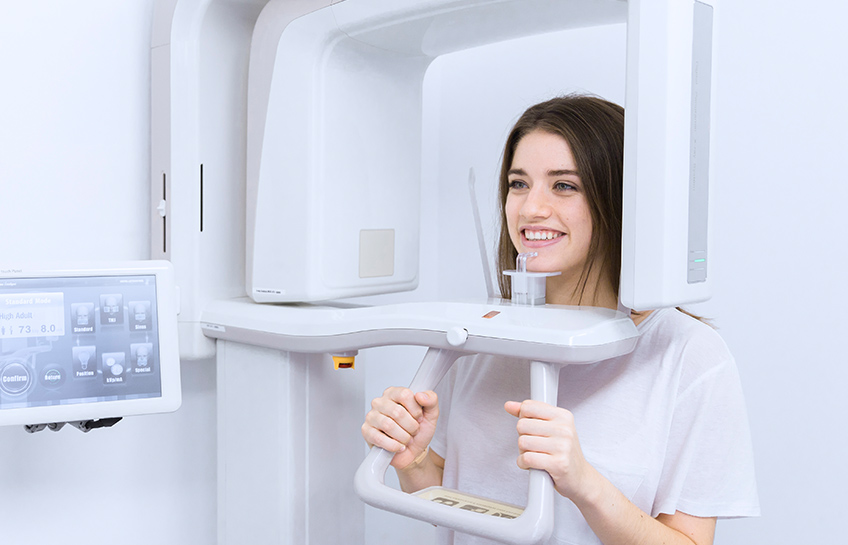
Cone-beam computed tomography scanning technology is an advanced form of 3D imaging that uses a special machine to produce detailed images of the teeth, soft tissues, and bone in the oral structures. Images produced with cone-beam CT scans provide excellent detail and resolution. These images are highly accurate and can be manipulated to help diagnose problems and make treatment planning easier.
At Dental Health Center, we use cone beam CT imaging technology for dental implant planning and other procedures such as root canals. With these precise 3D images, we can improve treatment planning and provide better treatment outcomes.

Common Applications of Cone Beam 3D Imaging
Dental Implant PlacementCBCT scans provide detailed three-dimensional images of the jawbone, allowing dentists to assess bone density, height, and width. This information is crucial for precise implant placement, determining the ideal size, length, and angulation of implants. CBCT imaging helps avoid vital structures, such as nerves or sinuses, and enhances the success and longevity of dental implant procedures.
Orthodontic Treatment PlanningCBCT imaging plays a significant role in orthodontic treatment planning. It provides detailed images of the teeth, jaws, and surrounding structures, allowing our dentists to analyze dental and skeletal relationships, identify impacted teeth, assess root positions, evaluate airway anatomy, and plan appropriate orthodontic interventions. CBCT imaging aids in precise diagnosis, treatment planning, and monitoring of the progress of orthodontic treatment.
CBCT scans are beneficial in endodontic procedures, especially in complex cases. They help diagnose complicated root canal anatomy, detect root fractures, assess the proximity of adjacent structures, and evaluate the success of root canal treatments. CBCT images provide detailed information that aids in making accurate treatment decisions.
Oral and Maxillofacial SurgeryCBCT images are extensively used in oral and maxillofacial surgery. They allow dentists to visualize the jaws, teeth, sinuses, nerve pathways, and anatomical structures in 3D. CBCT scans assist in planning procedures such as wisdom tooth extractions, dental extractions, corrective jaw surgery, bone grafting, and implant placement. Dentists use them to evaluate the relationship between the teeth, bones, and soft tissues for precise surgical planning and optimal outcomes.


The Benefits of Cone Beam 3D Imaging
CBCT imaging provides high-resolution, three-dimensional images of dental and maxillofacial structures. This allows for a comprehensive and detailed assessment of the teeth, jaws, bones, airways, and soft tissues. The ability to view structures from multiple angles helps dentists and specialists better understand the patient's anatomy.
CBCT technology provides additional diagnostic information that may not be visible with conventional two-dimensional dental imaging. It allows for detecting and evaluating dental and maxillofacial pathology, including cysts, tumors, infections, fractures, and other abnormalities. Detailed images enable early detection, accurate diagnosis, and appropriate treatment planning.
For more information, visit Dental Health Center at 56 Professional Plaza, Rexburg, ID 83440, or call (208) 356-9262. Our team of professional dentist in Rexburg, ID is committed to providing excellent service and efficient diagnosis and treatment for your oral health concerns.
